Staphylococcus aureus
| Staphylococcus aureus | |
 | |
| Systematik | |
|---|---|
| Domän | Bakterier Bacteria |
| Stam | Firmicutes |
| Klass | Bacilli |
| Ordning | Bacillales |
| Familj | Staphylococcaceae |
| Släkte | Stafylokocker Staphylococcus |
| Art | S. aureus |
| Vetenskapligt namn | |
| § Staphylococcus aureus | |
| Auktor | Rosenbach, 1884 |
Staphylococcus aureus (S. aureus), gula stafylokocker, är en bakterietyp som finns hos minst 50% av alla friska individer, de förekommer främst i näsöppningen. Trots det räknas de inte till normalfloran hos människan, utan de betraktas som patogena bakterier.[1] I de flesta fall orsakar bakterien ingen skada utan lever i symbios med människokroppen, oftast på huden eller i näsan. Staphylococcus aureus kan orsaka infektion i de flesta vävnader. Den är en mycket vanlig orsak till matförgiftning, variga hud- och sårinfektioner, skelettinfektioner och infektion i hjärtklaffarna, endokardit.[2]
Struktur och funktion
Bakterierna är runda, ungefär 1 mikrometer långa och grampositiva. Till skillnad från de flesta andra stafylokockarter är S. aureus koagulaspositiv[3]. Det innebär att den kan bilda ett enzym som heter koagulas, vilket kan koagulera blodplasma.[4]
Som sjukdomsorsak
Den kan också ge upphov till lunginflammation och mer sällan till hjärnhinneinflammation och urinvägsinfektion. I vissa fall kan infektionerna bli systemiska och i värsta fall ge upphov till det livshotande tillståndet septisk chock.[5] En ovanlig, men allvarlig stafylokocksjukdom är toxic shock syndrome som orsakas av ett gift som i vissa fall produceras av bakterien.[6]
MRSA
En undergrupp av de gula stafylokockerna kallas för MRSA (meticillinresistent staphylococcus aureus). Dessa är resistenta mot alla penicilliner, cefalosporiner och karbapenemer. I Sverige beräknas knappt en procent av S. aureus vara meticillinresistenta.[7] Det är internationellt sett mycket lågt;[8] i andra länder kan andelen MRSA uppgå till över 25 %.
Galleri
- Infektion av MRSA i huden som gett upphov till en abscess.
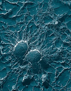
- Staphylococcus aureus i 20 000x förstoring.
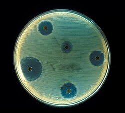
- Antibiotikatest av S. aureus.
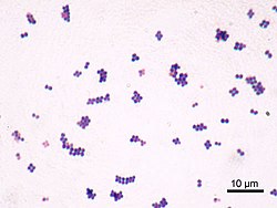
- Gramfärgning av S. aureus (grampositiv).
- Odling av S. aureus.
Referenser
Noter
- ^ Ericson, Elsy & Thomas (2009). Klinisk mikrobiologi. Läst 7 januari 2017
- ^ http://www.vardguiden.se/Article.asp?Articleid=3278/
- ^ Becker, Karsten; von Eiff, Christof (2011). ”19 Staphylococcus, Micrococcus, and other catalase-positive cocci”. i Versalovic James et al. (på engelska). Manual of Clinical Microbiology. 10th Edition. Washington DC, USA: ASM Press. sid. 310-11. ISBN 978-1-55581-463-2
- ^ ”MeSH Tree Location(s) for Coagulase”. Karolinska institutet. Arkiverad från originalet den 4 mars 2016. https://web.archive.org/web/20160304102212/http://mesh.kib.ki.se/swemesh/show.swemeshtree.cfm?Mesh_No=D12.776.097.181&tool=karolinska.
- ^ ”Sjukdomsinformation om meticillinresistenta gula stafylokocker (MRSA)”. Folkhälsomyndigheten. https://www.folkhalsomyndigheten.se/smittskydd-beredskap/smittsamma-sjukdomar/meticillinresistenta-gula-stafylokocker-mrsa/.
- ^ ”Sjukdomsinformation om toxic shock syndrome (TSS)”. Folkhälsomyndigheten. Arkiverad från originalet den 14 februari 2017. https://web.archive.org/web/20170214103200/https://www.folkhalsomyndigheten.se/smittskydd-beredskap/smittsamma-sjukdomar/toxic-shock-syndrome-tss-/. Läst 13 februari 2017.
- ^ ”Staphylococcus aureus”. Folkhälsomyndigheten. https://www.folkhalsomyndigheten.se/folkhalsorapportering-statistik/statistikdatabaser-och-visualisering/sjukdomsstatistik/staphylococcus-aureus/.
- ^ ”S.aureus - MRSA (methicillin- och multiresistens) och VRSA (vankomycinresistens)”. srga.org. Arkiverad från originalet den 17 mars 2010. https://web.archive.org/web/20100317122440/http://www.srga.org/mrb/mrbstaf.html.
Källor
Media som används på denna webbplats
Författare/Upphovsman: Y Tambe, Licens: CC BY-SA 3.0
microscopic image of Staphylococcus aureus (ATCC 25923). Gram staining, magnification:1,000.
An example of Petri dishes cultures of Staphylococcus aureus
Staphylococcus aureus - Antibiotics Test plate
This 2005 photograph (low resolution version) depicted a cutaneous abscess located on the hip of a prison inmate, which had begun to spontaneously drain, releasing its purulent contents. The abscess was caused by methicillin-resistant Staphylococcus aureus bacteria, referred to by the acronym MRSA.
S. aureus bacteria are amongst the populations of bacteria normally found existing on ones skin surface. However, over time, various populations of these bacteria have become resistant to a number of antibiotics, which makes them very difficult to fight when attempting to treat infections where MRSA bacteria are the responsible pathogens. These antibiotics include methicillin and other more common antibiotics such as oxacillin, penicillin and amoxicillin.
Staph infections, including MRSA, occur most frequently among persons in hospitals and healthcare facilities such as nursing homes and dialysis centers, who have weakened immune systems, however, the manifestation of MRSA infections that are acquired by otherwise healthy individuals, who have not been recently hospitalized, or had a medical procedure such as dialysis, or surgery, first began to emerged in the mid- to late-1990's. These infections in the community are usually manifested as minor skin infections such as pimples and boils. Transmission of MRSA has been reported most frequently in certain populations, e.g., children, sports participants, or as was the case here, jail inmates. (CDC) High resolution image (15.56 MB) available at PHIL.Bacterial cells of Staphylococcus aureus, which is one of the causal agents of mastitis in dairy cows. Its large capsule protects the organism from attack by the cow's immunological defenses. This image was taken at 50,000X magnification on a Transmission Electron Microscope of a heavy-metal coated replica of a freeze dried sample, (TEM) Plate #.9514. Sourced from Plate #.9513's information.
Under a very high magnification of 20,000x, this scanning electron micrograph (SEM) shows a strain of Staphylococcus aureus bacteria taken from a vancomycin intermediate resistant culture (VISA).
Under SEM, one can not tell the difference between bacteria that are susceptible, or multidrug resistant, but with transmission electron microscopy (TEM), VISA isolates exhibit a thickening in the cell wall that may attribute to their reduced susceptibility to vancomycin . See PHIL 11156 for a black and white version of this image. VISA and VRSA are specific types of antimicrobial-resistant staph bacteria. While most staph bacteria are susceptible to the antimicrobial agent vancomycin some have developed resistance. VISA and VRSA cannot be successfully treated with vancomycin because these organisms are no longer susceptibile to vancomycin. However, to date, all VISA and VRSA isolates have been susceptible to other Food and Drug Administration (FDA) approved drugs.
How do VISA and VRSA get their names?
Staph bacteria are classified as VISA or VRSA based on laboratory tests. Laboratories perform tests to determine if staph bacteria are resistant to antimicrobial agents that might be used for treatment of infections. For vancomycin and other antimicrobial agents, laboratories determine how much of the agent it requires to inhibit the growth of the organism in a test tube. The result of the test is usually expressed as a minimum inhibitory concentration (MIC) or the minimum amount of antimicrobial agent that inhibits bacterial growth in the test tube. Therefore, staph bacteria are classified as VISA if the MIC for vancomycin is 4-8µg/ml, and classified as VRSA if the vancomycin MIC is >16µg/ml.


