Sporangium
Ett sporangium kallas även sporgömme[1] och är det organ på en sporofyt som producerar sporer genom meios (reduktionsdelning). Varje cell som genomgår meios producerar fyra sporer (undantag finns, som hos Sporsäckssvampar). Ett sporangium kan, beroende på vad det sitter på för slags organism vara antingen homosporiskt eller heterosporiskt. Att ett sporangium (samt även den organism den sitter på) är homosporiskt innebär att både hanliga och honliga delar kommer att finnas på den organism som sporen gror ut till. Att ett sporangium är heterosporiskt innebär att den organism som sporen gror ut till antingen har bara hanliga, eller bara honliga delar.
Sporangier är vanligt förekommande i många grupper av organismer som inte är nära släkt. Bladmossor, Ormbunkar, Kitinsvampar, Rödalger, Slemsvampar och många fler. Även blomväxter och barrträd har sporangier även om de är små och väl dolda inne i kottar och blommor.
Ett sporangium kan vara skaftat eller oskaftat. Vissa ormbunkar har en annulus, ringformig bildning, runt utsidan av sporangiet som hjälper till att öppna det och sprida ut sporerna. Det är också vanligt med elatärer, sterila trådar, inne bland sporerna hos levermossor och fräkenväxter och fler grupper. Dessa elatärer är hygroskopiska och sporspridningen påverkas därför av luftfuktigheten.
Bilder
- (c) Lairich Rig, CC BY-SA 2.0
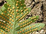
Sporangier på undersidan av stensötablad.
- (c) Lairich Rig, CC BY-SA 2.0
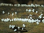
Sporangier hos en Myxomycetes, slemsvamp.
Referenser
- ^ http://www.ne.se/sporangium - från Nationalencyklopedin på nätet - http://www.ne.se - läst datum: 7 april 2014
| Den här artikeln behöver fler eller bättre källhänvisningar för att kunna verifieras. (2014-04) Åtgärda genom att lägga till pålitliga källor (gärna som fotnoter). Uppgifter utan källhänvisning kan ifrågasättas och tas bort utan att det behöver diskuteras på diskussionssidan. |
Media som används på denna webbplats
Författare/Upphovsman: Tkgd2007, Licens: CC BY-SA 3.0
A new incarnation of Image:Question_book-3.svg, which was uploaded by user AzaToth. This file is available on the English version of Wikipedia under the filename en:Image:Question book-new.svg
(c) Lairich Rig, CC BY-SA 2.0
Stalked slime mould fruiting bodies This stalked form is the most common type of spore-bearing structure among slime moulds. There would originally have been a single slime mould, in the form of a plasmodium (a slow-moving gelatinous mass of protoplasm), on or within this piece of wood; on fruiting, it divided up into many small units, each of which developed into one of these stalked sporangia (the plural of sporangium, the name for an individual spore-bearing structure); the spores develop in the upper part. In the species shown here, each of the sporangia was 2-3mm tall. At this stage, where the sporangia are still developing, and are gelatinous in consistency, very many species look greatly alike. Identification to species is only possible when the sporangia are mature, at which time they are no longer gelatinous, and they may be quite different in colour; they would, by then, have developed distinctive internal features that would allow the species to be identified after careful microscopic examination.
(c) Lairich Rig, CC BY-SA 2.0
Polypody (a fern) - the underside This species of fern was growing on a tree, beside one of the paths in the woodland walks at Ardardan.
There are three British species of Polypody: Southern (Polypodium cambricum), Common (P. vulgare), and Western (P. interjectum); there are also three hybrids (one for each of the three possible pairings of these species).
Visible here on the underside of the frond are the golden-coloured "sori"; a sorus is a cluster of tiny spore capsules ("sporangia"). The species shown here is not one of the hybrids; these cannot produce viable spores, and have only small purple sori. In addition, Southern Polypody does not occur in this region, so the fern found here is either Western or Common Polypody.
Distinguishing between these two species is not easy, since they are very similar. The features that can aid identification include the shape of the sori (either round or oval), and the shape of the finger-shaped leaflets, known as "pinnae", which may end in a sharp or a blunt tip, and which may have serrated edges or not. However, only a microscopic examination will give an identification with certainty. [See "The Fern Guide" by James Merryweather (3rd ed., 2007, Field Studies Council)]Författare/Upphovsman: Korporal, Licens: CC BY-SA 3.0
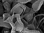
Scanning electron micrograph of fern sporangia in various stages of spore release.


