Röntgenundersökning


En röntgenundersökning, röntgendiagnostik eller röntgenologi är en undersökning av kroppens benstomme eller inre organ med hjälp av röntgenstrålning. Kroppen bestrålas med röntgenstrålning varvid olika vävnadstyper i kroppen släpper igenom olika mycket strålning till en bakomliggande fotografisk film eller digital detektor, så kallad attenuering. Efter framkallning framträder eventuella skador eller förändringar på röntgenfotot.
Undersökningsmetoden kallas vanligen slätröntgen eller konventionell röntgen. Undersökningen har också kallats skärmbildsundersökning. Röntgenfotot kallades tidigare också radiogram.
Diagnostik
Röntgenstrålarnas olika genomträngningsförmåga i mjuka respektive hårda vävnader ger en kontrastverkan som framträder på röntgenbilden, som är tillräcklig för att bedöma en bild av exempelvis lungorna eller ett benbrott. I andra fall måste man tillföra ett kontrastmedel, exempelvis en uppslamning av bariumsulfat vid undersökning av magtarmkanalen eller jodkontrast när man skall undersöka gallvägar, urinvägar, livmoderhåla och äggledare eller blodkärl.[1]
Digitalisering
Under de senare åren har man mer och mer övergått till digitala bilder, antingen från direktdigitala detektorer eller bildplattor. Den direktdigitala detektorn sitter antingen fast i undersökningsstativet, till exempel ett bord, eller är ansluten med en kabel till den bildinsamlande datorn. Fördelen är att man får en bild presenterad mycket snabbt, inom cirka tre sekunder samt att man inte behöver bära omkring kassetter som skall "framkallas".
Bildplattetekniken bygger på att man ersätter filmen med en strålningskänslig platta i en speciell sorts kassett. Kassetten kan då användas precis som man gjorde tidigare med filmkassetter och man behöver därmed inte bygga om undersökningsrummen.
För hantering av de digitala bilderna används regelmässigt standarden DICOM.
En modern utveckling är datortomografi.
Se även
Referenser
- ^ Bra Böckers lexikon, 1979.
|
Media som används på denna webbplats
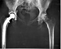
An A-P X-ray of a pelvis showing a total hip joint replacement. The right hip joint (on the left in the photograph) has been replaced. A metal prostheses is cemented in the top of the right femur and the head of the femur has been replaced by the rounded head of the prosthesis. A white plastic cup is cemented into the acetabulum to complete the two surfaces of the artificial "ball and socket" joint.
Although not the case here, hip prostheses can also be made of a ceramic material which rarely wears out during the patient's lifetime. During the operation, a bonding cement is used to fix the metal prostheses into the shaft of the femur and the plastic cup to the acetabulum (socket in the hip bone). One of the leading reasons for hip replacement is osteoarthritis of the hip joint in which virtually all of the cartilage around the top of the femur bone deteriorates due to wear over time, leaving a grinding bone-on-bone situation with the bone surfaces becoming roughened leading to pain and stiffness. Narrowing of joint space (the space between the acetabulum and the head of the femur) is also a feature of osteoarthritis. There may be other changes which are not entirely clear on this A-P X-ray, which probably should be reported together with lateral X-rays of the hips, or with modern computerised imaging techniques.
Keywords: total hip replacement, prosthesis, osteoarthritis, X-ray.Författare/Upphovsman: Setzner1337, Licens: CC0
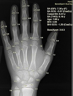
X-ray of the hand of a 7.60 year old female, with automatic calculation of bone age by BoneXpert. The bone age is 7.38 by the Greulich and Pyle algorithm, and 8.16 by the second Tanner Whitehouse algorithm.

