Portal:Medicin
| Denna sida är museimärkt och hålls inte längre uppdaterad. Den behålls främst av historiska skäl. Se eventuellt diskussionssidan för mer information. |
| ||||||||||||||||||||||||||||||||||||||||||||
|
|
| ||||||||||||||||||||||||||||||||||||||||||
Arkitektur · Azerbajdzjan · Buddhism · Djur · EU · Helsingborg · Hästsport · Ryssland · Stockholm | ||||||||||||||||||||||||||||||||||||||||||||
Media som används på denna webbplats
Museum icon
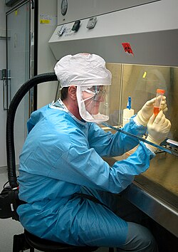
This 2005 photograph depicts Dr Terrence Tumpey, one of the Centers for Disease Control and Prevention’s staff microbiologists, examining reconstructed 1918 Pandemic Influenza Virus contained within a calibrated vial with a supernatant culture medium.
The photo is taken in a Biosafety Level 3-enhanced laboratory setting, where scientists work beneath a flow hood, where air outside the hood is pulled into the hood's confines and is then filtered of any pathogens before being recirculated inside the self-contained laboratory atmosphere.
Dr. Tumpey was chosen to be the only person authorized to work on the 1918 virus, and then only under biosecurity level 3 enhanced (BSL-3E) precautions. For example, to reduce risk to colleagues, Dr. Tumpey worked alone after normal agency hours. His health was continually monitored and he took influenza antiviral drugs preventively as a precaution in case he was exposed to infectious virus. [1]
The 1918 virus was recreated in order to identify the characteristics that made this organism such a deadly pathogen. Research efforts such as this, enables researchers to develop new vaccines and treatments for future pandemic influenza viruses.
The 1918 Spanish flu epidemic was caused by an influenza A (H1N1) virus, killing more than 500,000 people in the United States, and up to 50 million worldwide. The possible source was a newly emerged virus from a swine or an avian host of a mutated H1N1 virus. Many people died within the first few days after infection, and others died of complications later. Nearly half of those who died were young, healthy adults. Influenza A (H1N1) viruses still circulate today after being introduced again into the human population in the 1970s.Författare/Upphovsman: SOLEIL, Licens: CC BY-SA 3.0
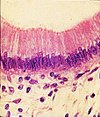
Simple columnar epithelium. HyE.
Författare/Upphovsman: James Heilman, MD, Licens: CC BY-SA 3.0
A cat scan demonstrating acute appendicitis (note the appendix has a diameter of 17.1mm)
Författare/Upphovsman: Intropin, Licens: CC BY 3.0
Diphenhydramine HCl preparation, single dose vial for IV administration.
Nurse putting drops in new-born baby's eyes. Interior. New Orleans, 1936. WPA Public Nursing Project.
(c) Bundesarchiv, Bild 183-J0603-0007-001 / CC-BY-SA 3.0
Författare/Upphovsman: Kieran Maher and Dirk Hünniger, Licens: CC BY-SA 3.0
B-mode image from a patient's liver scan.
This colorized version of PHIL 232 depicts a scanning electron micrograph (SEM) of a number of Pseudomonas aeruginosa bacteria.
Författare/Upphovsman: Wei-Chung Allen Lee, Hayden Huang, Guoping Feng, Joshua R. Sanes, Emery N. Brown, Peter T. So, Elly Nedivi, Licens: CC BY 2.5
After the original figure legend: Coronal section containing the chronically imaged pyramidal neuron “dow” (visualized by green GFP) does not stain for GABA (visualized by antibody staining in red). Confocal image stack, overlay of GFP and GABA channels. Scale bar: 100 μm
An anatomical study of the principal organs and the arterial system of a female torso, pricked for transfer.
Författare/Upphovsman: P Thomas et al., Licens: CC BY 2.0
Axial section computed tomography scan demonstrating
hyperdensity in the left sigmoid sinusInfo icon with hourglass
Författare/Upphovsman: Clinica e centro de pesquisa em reprodução humana Roger Abdelmassih, Licens: Attribution
Inicío da injecção do espermatozóide no óvulo
Gastroscopy image of esophageal varices with prominent red wale spots.
Författare/Upphovsman: Liem T. Bui-Mansfield et al, Licens: CC BY 2.0
Coronal CT scan shows osteochondral lesion with loose body (arrow) in medial aspect of tibial plafond.
Författare/Upphovsman: Hospital, Licens: CC BY-SA 3.0
Andningsballong (Rubens Blåsa, AMBU-bag), Revivator. Används för manuell vantilering av patient vid andningssvårigheter eller andningsstillestånd.
Författare/Upphovsman: James Heilman, MD, Licens: CC BY-SA 3.0
A small bone spur as marked by the arrow.
Författare/Upphovsman:
- Original: CatherinMunro
- härlett verk: Rehua
Rod of Asclepius
Författare/Upphovsman: Yohan euan o4, Licens: CC BY-SA 3.0
Beecham's All in One
Författare/Upphovsman: Rakesh Ahuja, MD, Licens: CC BY-SA 3.0
Cataract in Human Eye


