Merkelcellskarcinom
| Merkelcellskarcinom | |
 Mikrografi av ett merkelcellskarcinom. H&E färgning. | |
| Klassifikation och externa resurser | |
|---|---|
| ICD-10 | C44 (ILDS C44.L44) |
| ICD-9 | 209.31 - 209.36 |
| ICD-O | M8247/3 |
| DiseasesDB | 31386 |
| eMedicine | DERM/262 ent/714 |
| MeSH | svensk engelsk |
Merkelcellskarcinom är en ovanlig men aggressiv form av hudcancer. Det finns en koppling mellan denna hudtumör och ljus hudtyp, solexponering, hög ålder och nedsatt immunförsvar.[1] Mer än hälften av alla merkelcellskarcinom är belägna i huvud- och halsregionen vilket talar för att solens UV-strålning är av stor betydelse vid denna diagnos.[2] År 2008 upptäcktes att merkelcellskarcinom har en klar koppling till ett virus som döpts till Merkel Cell Polyomavirus.[3]
Trots att merkelcellskarcinom är en ovanlig form av hudcancer så har förekomsten hos Sveriges befolkning fördubblats de senaste 20 åren.[2] MCC börjar ofta med en plötslig tillväxt av en röd, rosa eller rödviolett samt relativt hård upphöjning på eller under huden. Tumören växer snabbt till en knuta. Blödning kan ske i mer avancerade stadier. Tidig upptäckt och behandling med kirurgi och/eller strålbehandling är viktig.[4] Risken för återväxt av tumören i huden, spridning till lymfkörtlar eller andra organ är relativt hög.
Referenser
- ^ Heath M, Jaimes N, Lemos B, Mostaghimi A, Wang LC, Penas PF, et al. Clinical characteristics of Merkel cell carcinoma at diagnosis in 195 patients: the AEIOU features. Journal of the American Academy of Dermatology. 2008; 58: 375-81.
- ^ [a b] Zaar O, Gillstedt M, Lindelöf B, Wennberg-Larkö AM, Paoli J. Merkel cell carcinoma incidence is increasing in Sweden. J Eur Acad Dermatol Venereol. 2016 May 2. doi: 10.1111/jdv.13698. [Epub ahead of print]
- ^ Feng H, Shuda M, Chang Y, Moore PS. Clonal integration of a polyomavirus in human Merkel cell carcinoma. Science. 2008; 319: 1096-100.
- ^ ”Merkelcellskarcinom”. www.internetmedicin.se. http://www.internetmedicin.se/page.aspx?id=5697. Läst 12 juli 2016.
Galleri av bilder

Merkelcellskarcinom på underarm

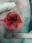
Stor merkel celltumörprov
Merkelcellskarcinom dissekeras under operation

Mikrofotografi av merkelcellskarcinom
Media som används på denna webbplats

Författare/Upphovsman: Ed Uthman from Houston, TX, USA, Licens: CC BY-SA 2.0
This large tumor presented as a protuberant mass on the buttock of a 45-year-old woman. Microscopically it showed sheets of undifferentiated cells with high nuclear/cytoplasmic ratio, a high mitotic rate, a occasional inconspicuous nucleoli.
Immunostains showed strong dot-like and diffuse cytoplasmic positivity for cytokeratins 20, CAM5.2, and AE1-AE3, as well as granular staining for synaptophysin and chromogranin. There were negative reactions for cytokeratin 7, gross cystic disease fluid protein 15, melan-A, S-100 protein, and thyroid transcription factor 1-alpha.
Even though the histologic features and immunostain profile was characteristic of a primary skin tumor, the presentation is atypical. Usually, Merkel cell carcinoma presents on the sun-exposed areas of elderly individuals. Therefore, the pathologist recommended workup for the possibility of another primary site, including lung, female genital tract, salivary gland, and other cutaneous site.
Författare/Upphovsman: Nakos histopathology, Licens: CC BY-SA 4.0
During macroscopic examination
Författare/Upphovsman: Nephron, Licens: CC BY-SA 3.0
Very high magnification micrograph of Merkel cell carcinoma, abbreviated MCC. H&E stain.
MCCs are aggressive neuroendocrine tumour of the skin. They can be considered a small round cell tumour.
Most MCCs are caused by a virus aptly named Merkel cell polyomavirus.[1]
Related images
-
 Intermed. mag.
 -
 High mag.
 -
 Very high mag.
 -
 Very high mag. (cropped)


References
- ↑ (Feb 2008). "Clonal integration of a polyomavirus in human Merkel cell carcinoma.". Science 319 (5866): 1096-100. DOI:10.1126/science.1152586.

Författare/Upphovsman: Patrick S. Moore, Licens: CC BY-SA 4.0
Photomicrograph of Merkel cell carcinoma infiltrating the skin (arrow). Tumor cell nuclei are stained brown by an antibody to the Merkel cell polyomavirus large T antigen.

Författare/Upphovsman: John Paoli (GU), Licens: CC BY-SA 4.0
Merkelcellskarcinom på underarm.