Mellankropp
| Den här artikeln behöver källhänvisningar för att kunna verifieras. (2021-09) Åtgärda genom att lägga till pålitliga källor (gärna som fotnoter). Uppgifter utan källhänvisning kan ifrågasättas och tas bort utan att det behöver diskuteras på diskussionssidan. |
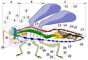
Mellankropp, eller thorax, är en av de tre övergripande delar som kroppen av en insekt anatomiskt uppdelas i. Det är den delen av kroppen som är mellan huvudet och bakkroppen. På mellankroppen sitter benen och vingarna.
Mellankroppen är uppdelad i tre segment, i ordningsföljd från det första till det sista benämnda prothorax, mesothorax och metathorax. Till varje segment hör ett par ben. De två sista segmenten bär hos bevingade insekter vardera även ett par vingar (som kan vara rudimentära som hos boklöss eller ombildade som svängkolvarna hos tvåvingar). Segmentens yttre hudskelett har vart och ett en ryggplåt (tergit) och en bukplåt (sternit). På sidorna finns dessutom sidoplåtar (pleuriter). Dessa plåtar kallas med ett gemensamt namn skleriter.
Hos trollsländor är segmenten mesothorax och metathorax sammanfogade och benämns synthorax.
Media som används på denna webbplats
Författare/Upphovsman: Tkgd2007, Licens: CC BY-SA 3.0
A new incarnation of Image:Question_book-3.svg, which was uploaded by user AzaToth. This file is available on the English version of Wikipedia under the filename en:Image:Question book-new.svg
Författare/Upphovsman: Internet Archive Book Images, Licens: No restrictions
Identifier: entomologywit00fols (find matches)
Title: Entomology : with special reference to its biological and economic aspects
Year: 1906 (1900s)
Authors: Folsom, Justus Watson, 1871-1936
Subjects: Entomology
Publisher: Philadelphia : P. Blakiston's Son
Contributing Library: Robarts - University of Toronto
Digitizing Sponsor: University of Toronto
View Book Page: Book Viewer
About This Book: Catalog Entry
View All Images: All Images From Book
Click here to view book online to see this illustration in context in a browseable online version of this book.
Text Appearing Before Image:
Tracheal system of an insect, a, an-tenna; b, brain; /, leg; 11, nerve cord;p, palpus; s, spiracle; st, spiracular, orstigmatal, branch; t, main tracheal trunk;V, ventral branch; is, visceral branch.—After KoLBE. ANATOMY AND PHYSIOLOGY 133 9. Respiratory SystemIn insects, as contrasted with vertebrates, the air itself isconveyed to the remotest tissues by means of an elaborate sys-tem of branching air-tuljes, or iracJiccv, which receive airthrough paired segmentally-arranged spiracles. Each spiracleis commonly the mouth of a short tube which opens into amain tracheal trunk (Fig. 167) extending along the side of
Text Appearing After Image:
Diagrammatic cross section of the thorax of an insect, a, alimentarj canal; d,dorsal vessel; g, ganglion; s, spiracle; w, wing; /, dorsal tracheal branch; 3, visceralbranch; 3, ventral branch. the body. From the two main trunks branches are sent whichdivide and subdivide until they become extremelv delicatetubes, which penetrate even between muscle fibers, between theommatidia of the compound eyes and possibly enter cells. Inmost cases each main longitudinal trunk gives off in each seg-ment (Fig. 168) three large branches: (i) an upper, or dor-sal, branch, which goes to the dorsal muscles; (2) a middle,or visceral, branch, which supplies the alimentary tract and thereproductive organs; (3) a lower, or ventral, branch, whichpertains to the ventral ganglia and muscles. In many swiftly-flying insects (dragon flies, beetles, moths,flies and bees) there occur tracheal pockets, or air-sacs, which 134 ENTOMOLOGY Fig. 169. were formerly and erroneously supposed to diminish theweight of the in
Note About Images
Författare/Upphovsman: Piotr Jaworski, PioM, Licens: CC BY-SA 3.0
Anatomic Diagram of an insect


