Kopplingssvampar
| Den här artikeln eller det här avsnittet innehåller inaktuella uppgifter och behöver uppdateras. (2023-04) Motivering: se i Dyntaxa, ej längre gällande taxon Hjälp gärna Wikipedia att åtgärda problemet genom att redigera artikeln eller diskutera saken på diskussionssidan. |
| Kopplingssvampar | |
 Närbild på ett Phycomyces sporangium. | |
| Systematik | |
|---|---|
| Domän | Eukaryoter Eukaryota |
| Rike | Svampar Fungi |
| Division | Kopplingssvampar Zygomycota |
| Vetenskapligt namn | |
| § Zygomycota | |
| Auktor | Moreau 1954 |
| Ordningar | |
| |
Kopplingssvampar (Zygomycota eller Zygomyceter) är en division i svampriket med cirka 1060 arter i världen. Till divisionen hör bland annat Mucor och Rhizopus som i dagligt tal kallas kulmögel. Både Mucor och Rhizopus kan finnas på frukt och spannmål. De bryter ner livsmedlet utan att tillverka toxiner.[1]
Fylogenetik
Divisionen Zygomycota placeras generellt sett nära basen på svamparnas fylogenetiska träd-struktur, eftersom de divergerade från andra svampar efter Chytridiomycota.
Molekylär fylogenetik avslöjar att kopplings-svamparna utgör en polyfyletisk grupp, vilken kan komma att splittras i flera nya divisioner.[2][3][4]
Ordningen Glomales förflyttades år 2001 och upphöjdes till division (eller phylum) Glomeromycota.[5][6]
Bildgalleri
Referenser
- ^ Marie Widén, Björn Widén (red), Botanik: Systematik, Evolution, Mångfald. 2008. ISBN 978-91-44-04304-3
- ^ Hibbett DS, Binder M, Bischoff JF, et al. (maj 2007). A higher-level phylogenetic classification of the Fungi. Mycol. Res. 111 (Pt 5): 509–47. doi:10.1016/j.mycres.2007.03.004. PMID 17572334.
- ^ Bisby F.A., Roskov Y.R., Orrell T.M., Nicolson D., Paglinawan L.E., Bailly N., Kirk P.M., Bourgoin T., Baillargeon G., Ouvrard D. (red.) (5 september 2011). ”Species 2000 & ITIS Catalogue of Life: 2011 Annual Checklist.”. Species 2000: Reading, UK. http://www.catalogueoflife.org/annual-checklist/2011/search/all/key/zygomycota/match/1. Läst 24 september 2012.
- ^ Dyntaxa Zygomycota
- ^ Tree of Life web project: Zygomycota. Läst 2009-03-07.
- ^ Schüßler A., Schwarzott D., Walker C. (2001). A new fungal phylum, the Glycomycota: phylogeny and evolution. Mycol. Res. 105 (12): 1413–1421. doi:10.1017/S0953756201005196.
Media som används på denna webbplats
An outdated clock with a serious icon
Författare/Upphovsman: Muslihfsu, Licens: CC BY 3.0
Figure5. Lipid globules and protein crystals in sporangiophore. The numbers indicate the following: 1. Lipid globules; 2. Protein crystals; 3. Vacuolar transepts; 4. Central vacuole
Författare/Upphovsman: Maya and Rike, Licens: CC BY 3.0
Figure1. Cell wall structure of Fungi
Författare/Upphovsman: TheAlphaWolf, Licens: CC BY-SA 3.0
Closeup of the Phycomyces sporangium.
Författare/Upphovsman: Muslihfsu, Licens: CC BY 3.0
Fig. 2: Sporangium. *occurs only in some genera
Författare/Upphovsman: Muslihfsu, Licens: CC BY 3.0
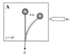
Figure4. Bending angle in vertical sporangiophore: the gravitropic stimulus increases in proportion to the increasing bending angle
Författare/Upphovsman: Muslihfsu, Licens: CC BY 3.0
Fig.3 Postulated biosynthesis of trisporic acid B
Författare/Upphovsman: Muslihfsu, Licens: CC BY 3.0
Figure7. Zygomycetes on Sub-media, in light and non-light condition
Författare/Upphovsman: Muslihfsu, Licens: CC BY 3.0
Figure8. Zygomycetes on solid media.
Författare/Upphovsman: Muslihfsu, Licens: CC BY 3.0
Figure6. Phylogenetic tree of True Fungi.


