Histoplasma capsulatum
| Den här artikeln har skapats av Lsjbot, ett program (en robot) för automatisk redigering. (2012-10) Artikeln kan innehålla fakta- eller språkfel, eller ett märkligt urval av fakta, källor eller bilder. Mallen kan avlägsnas efter en kontroll av innehållet (vidare information) |
| Histoplasma capsulatum | |
 | |
| Systematik | |
|---|---|
| Domän | Eukaryoter Eukaryota |
| Rike | Svampar Fungi |
| Division | Sporsäcksvampar Ascomycota |
| Klass | Eurotiomycetes |
| Ordning | Onygenales |
| Familj | Ajellomycetaceae |
| Släkte | Histoplasma |
| Art | Histoplasma capsulatum |
| Vetenskapligt namn | |
| § Histoplasma capsulatum | |
| Auktor | Darling 1906 |
| Synonymer | |
| Posadasia capsulata (Darling) M. Moore 1934[1] Torulopsis capsulata (Darling) Castell. & Jacono 1933[1] Torulopsis capsulata (Darling) F.P. Almeida 1933[1] Grubyella farcinimosa (Rivolta) M. Ota 1925[1] Cryptococcus capsulatus (Darling) Castell. & Chalm. 1919[2] Histoplasma capsulatum var. capsulatum Darling 1906[3] | |
Histoplasma capsulatum är en svampart[3] som orsakar sjukdomen Histoplasmos. Svampen beskrevs av Darling 1906. Histoplasma capsulatum ingår i släktet Histoplasma och familjen Ajellomycetaceae.[4][5] Inga underarter finns listade i Catalogue of Life.[4]
Källor
- ^ [a b c d] ”CABI databases”. http://www.speciesfungorum.org. Läst 24 januari 2013.
- ^ Castell. & Chalm. (1919) , In: Manual of tropical medicine (London):1076
- ^ [a b] Darling (1906) , In: Journal of the American Medical Association 46:1285
- ^ [a b] Bisby F.A., Roskov Y.R., Orrell T.M., Nicolson D., Paglinawan L.E., Bailly N., Kirk P.M., Bourgoin T., Baillargeon G., Ouvrard D. (red.) (1 september 2011). ”Species 2000 & ITIS Catalogue of Life: 2011 Annual Checklist.”. Species 2000: Reading, UK. http://www.catalogueoflife.org/annual-checklist/2011/search/all/key/histoplasma+capsulatum/match/1. Läst 24 september 2012.
- ^ Species Fungorum. Kirk P.M., 2010-11-23
 Wikimedia Commons har media som rör Histoplasma capsulatum.
Media som används på denna webbplats
Robot icon
This chest film shows diffuse pulmonary infiltration due to acute pulmonary histoplasmosis caused by H. capsulatum.
Se observa la distribución de la histoplasmosis alrededor del mundo
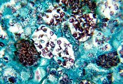
This map appears to be inaccurate: see, e.g. the Journal of the Venezuelan Society of Microbiology for a map that reflects a totally different distribution in S. America.Note the histopathologic changes seen in histoplasmosis due to Histoplasma capsulatum var. duboisii. Note the presence of typical yeast cells, some of which are undergoing replication by “budding”. Histoplasmosis can be confined to the lungs, or become systemically disseminated, thereby, producing a fatal outcome. Methenamine silver stain was used here.
This patient presented with a skin lesion on his upper lip. The ulcer was originally thought to be due to a syphilitic infection, but later, after laboratory testing, was diagnosed as histoplasmosis, due to the fungus Histoplasma capsulatum. Skin lesions such as this can be a manifestation of “disseminated histoplasmosis”, referring to the spread of the fungal pathogen, H. capsulatum, throughout the body including the skin, bone marrow, brain and other organs. Disseminated histoplasmosis is most common amongst immunosuppressed people, such as those with AIDS.
Photograph of an eye with Presumed ocular histoplasmosis syndrome
Författare/Upphovsman: Yale Rosen, Licens: CC BY-SA 2.0
Histoplasmosis - GMS stain
Small yeast forms with budding seen at arrows.Författare/Upphovsman: Yale Rosen, Licens: CC BY-SA 2.0
Histoplasmosis
Old necrotizing fibrocalcific granulomasID#: 4023 Description: This photomicrograph shows two tuberculate macroconida of the Jamaican isolate of Histoplasma capsulatum.
Histoplasma capsulatum grows in soil, and material contaminated with bat or bird droppings. Spores become airborne when contaminated soil is disturbed, and breathing the spores causes histoplasmosis, a disease not transmitted from person to person.
Content Providers(s): CDC/Dr. Libero Ajello Creation Date: 1968
Copyright Restrictions: None - This image is in the public domain and thus free of any copyright restrictions. As a matter of courtesy we request that the content provider be credited and notified in any public or private usage of this image.Note the histopathologic changes seen in histoplasmosis due to Histoplasma capsulatum using methenamine silver stain. Note the presence of typical yeast cells, some of which are undergoing replication by “budding”. Histoplasmosis can be confined to the lungs, or become systemically disseminated, thereby, producing a fatal outcome.


