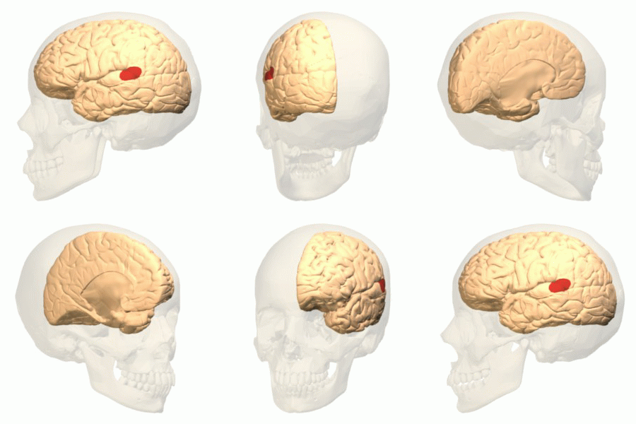
Wernicke's area animation
Författare/Upphovsman:
Polygon data were generated by Database Center for Life Science(DBCLS)[2].
Tillskrivning:
Bilden är taggad "Attribution Required" men ingen tillskrivningsinformation lämnades. Attributionsparametern utelämnades troligen när MediaWiki-mallen användes för CC-BY-licenserna. Författare och upphovsmän hittar ett exempel för korrekt användning av mallarna här.
Kreditera:
Polygon data are from BodyParts3D[1]
Kort länk:
Källa:
Upplösning:
300 x 300 Pixel (2085080 Bytes)
Beskrivning:
Wernicke's area (shown in red).
Colored region is posterior section of the superior temporal gyrus (pSTG) of the left cerebral hemisphere. Though this region is generally treated as Wernicke's area, there are many researches and discussions about its exact size and anatomical boundaries.
Colored region is posterior section of the superior temporal gyrus (pSTG) of the left cerebral hemisphere. Though this region is generally treated as Wernicke's area, there are many researches and discussions about its exact size and anatomical boundaries.
- Wise RJ, Scott SK, Blank SC, Mummery CJ, Murphy K, Warburton EA. "Separate neural subsystems within 'Wernicke's area'. " Brain: 2001, 124(Pt 1);83-95 PMID 11133789
- Tomoo Inubushi, Kuniyoshi Sakai (2014) "Language center" Brain Science Dictionary http://bsd.neuroinf.jp/wiki/%E8%A8%80%E8%AA%9E%E4%B8%AD%E6%9E%A2 (in Japanese)
Licens:
Licensvillkor:
Creative Commons Attribution-Share Alike 2.1 jp
Mer information om licensen för bilden finns här. Senaste uppdateringen: Fri, 11 Oct 2024 11:37:53 GMT
