Myxomatous aortic valve
Författare/Upphovsman:
Tillskrivning:
Bilden är taggad "Attribution Required" men ingen tillskrivningsinformation lämnades. Attributionsparametern utelämnades troligen när MediaWiki-mallen användes för CC-BY-licenserna. Författare och upphovsmän hittar ett exempel för korrekt användning av mallarna här.
Kreditera:
Eget arbete
Kort länk:
Källa:
Upplösning:
2048 x 1536 Pixel (349803 Bytes)
Beskrivning:
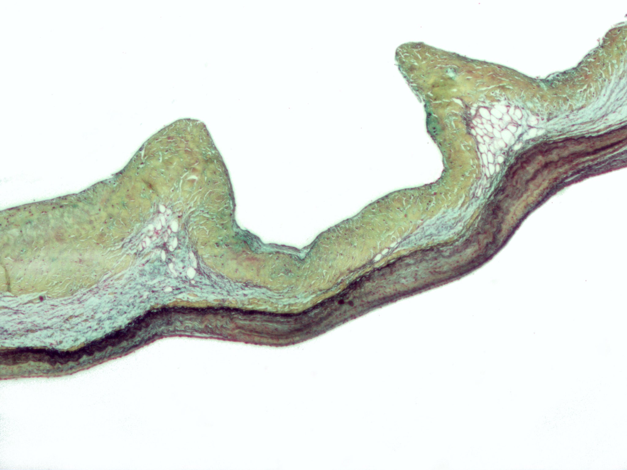
Micrograph of myxomatous degeneration of the aortic valve. Surgical specimen. Movat's stain (Black = nuclei, elastic fibres. Yellow = collagen, reticular fibers. Blue =
ground substance, mucin. Bright red = Fibrin. Red = muscle.)
In myxomatous degeneration, the ventricularis layer (composed primarily of elastic tissue) is thinned and the spongiosa layer (composed of loose connective tissue) is thickened.
On the image, the fibrosa layer (composed of collagen) is on the top, the thickened spongiosa layer below it and the ventricularis layer (made of elastic tissue) at the bottom.
The ventricularis layer, as the name may suggest, is closest to the (left) ventricle. The fibrosa layer is closest to the sinus of valsalva.
See also
- Marfan's syndrome - a condition, due to a defect in fibrillin (an essential component of elastic fibers), in which myxomatous degeneration is common.
Licens:
Licensvillkor:
Creative Commons Attribution-Share Alike 3.0
Mer information om licensen för bilden finns här. Senaste uppdateringen: Wed, 03 Apr 2024 22:49:01 GMT