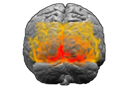
Brodmann areas 17 18 19
Författare/Upphovsman:
The original uploader was Washington irving på engelska Wikipedia.
Tillskrivning:
Bilden är taggad "Attribution Required" men ingen tillskrivningsinformation lämnades. Attributionsparametern utelämnades troligen när MediaWiki-mallen användes för CC-BY-licenserna. Författare och upphovsmän hittar ett exempel för korrekt användning av mallarna här.
Kreditera:
These images were created using Blender and Matlab.
. Originally from en.wikipedia; description page is/was here.
Kort länk:
Källa:
Upplösning:
256 x 192 Pixel (40769 Bytes)
Beskrivning:
Brodmann areas 17, 18 and 19. BA 17 is shown in red. BA 18 is orange. BA 19 is yellow. This is a rear view of the brain. Much of BA 17 is hidden from view on the medial surface (between the hemispheres), on the ventral bank of the calcarine sulcus (check this last point). The brain's surface is extracted from structural en:MRI data (Wellcome Dept. Imaging Neuroscience, UCL, UK). The Brodmann Area data is based on information from the online Talairach demon (an electronic version of Talairach and Tournoux, 1988).
Licens:
Licensvillkor:
Creative Commons Attribution-Share Alike 3.0
Mer information om licensen för bilden finns här. Senaste uppdateringen: Thu, 05 Sep 2024 12:51:04 GMT
